
male > females
average age at presentation is 35 to 50
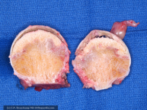
bilateral hips involved 80% of the time
Risk factors
direct causes
irradiation
trauma ( neck fractures)
hematologic diseases (leukemia, lymphoma)
dysbaric disorders (decompression sickness, "the bends") - Caisson disease
marrow-replacing diseases (e.g. Gaucher's disease)
sickle cell disease
indirect causes
alcoholism
hypercoagulable states
steroids (either endogenous or exogenous)
systemic lupus erythematosus (SLE)
transplant patient
virus (CMV, hepatitis, HIV, rubella, rubeola, varicella)
protease inhibitors (type of HIV medication)
idiopathic

Steinberg classification ( modification of Ficat classification)

Staeg 1

Stage 2

Stage 3

Stage 4

Stage 5

Stage 6
Symptoms
insidious onset of pain
pain with stairs, inclines, and impact
pain common in anterior hip
Physical exam
mostly normal initially
advanced stages similar to hip OA (limited motion, particularly internal rotation)
Imaging
Xrays ( bilateral): AP + L + frog leg
MRI:
double density appearance
T1: dark (low intensity band)
T2: focal brightness (marrow edema)
order when radiographs negative and osteonecrosis still suspected
presence of bone marrow edema on MRI is predicitve of worsening pain and future progression of disease
Nonoperative Treatment
bisphosphonates
indicated for precollapse AVN (Ficat stages 0-II)
prevents collapse ??
Surgical treatment
core decompression with or without bone grafting
indications
for early AVN, before subchondral collapse occurs
reversible etiology
Total hip arthroplasty

