Proximal femur fractures
Femoral neck fractures
Increasingly common due to aging population
Associated with high mortality rate (~25-30% at one year): pre-injury mobility is the most significant determinant for post-operative survival
Clinical prensetation
Pain in the groin or pain referred along the medial side of the thigh and knee
Leg in external rotation and abduction, with shortening ( if displaced)
Classification: Garden (for displacement) and Pauwels (for instbaility)


Garden Fracture
Can J Surg. 2003 Apr; 46(2): 147.

Pauwels classification
Journal of orthopaedic trauma. 15. 358-60.
Imaging
Xrays
Scan / MRI for occulte fracture ( not seen on xrays)

Xray showing a femoral neck fracture

CT scan was needed to confirm the femoral neck fracture

Occulte fracture shown only on MRI
Acta Orthopeadica. 2005 Aug; 76 (4): 524-530
Treatment : Surgery is required ( unless the patient does not ambulate or is at extreme risk for surgical intervention). Many surgical interventions exist according to the type of the fracture
Osteosynthesis :
Cannulated screws
Nondisplaced transcervical fx
Garden I or II in the physiologically elderly
Displaced transcervical fx in young patient (anatomical reduction is urgently needed)
Dynamic hip screw
Basicervical fracture
Vertical fracture pattern in a young patient
Arthroplasty
Total hip replacement vs hemiarthroplasty

Screws

Dynamic hip screw

Hemiarthroplasty

Total hip arthroplasty
Intertrochanteric Fractures
Extracapsular fractures of the proximal femur between the greater and lesser trochanters
Nonunion and malunion rates are low
Classified as stable and unstable
Stable: intact posteromedial cortex
Unstable:
comminution of the posteromedial cortex
thinner lateral wall thickness: <20.5 mm suggests risk of postoperative lateral wall fracture

The lateral wall thickness
Hsu, C.-E., Shih, C.-M., Wang, C.-C., & Huang, K.-C. (2013)

Xray showing an intertrochanteric fracture
Clinical presentation: painful, shortened, externally rotated lower extremity
Imaging : Xrays . CT scan/MRI are requested only for occulte fractures
Treatment:
Non operative (nonambulatory patients or at high risk)
Surgery
Dynamic hip screw : for stable cases
Intramedullary nail (for stable & unstable cases)
Arthroplasty
Severely comminuted fractures
Preexisting symptomatic degenerative arthritis
Osteoporotic bone that is unlikely to hold internal fixation
Salvage for failed internal fixation

DHS

Prosthesis
Intramedullary nail
Subtrochanteric fractures
Subtrochanteric typically defined as area from lesser trochanter to 5cm distal
Proximal fragment is in abduction, flexion and external rotation
Distal fragment is in adduction and shortened

Types of subtrochanteric fractures
Rule out pathologic or atypical femur fracture
denosumab or bisphosphonate use, particularly alendronate, can be risk factor
on Xrays: Transverse fracture line, Cortical thickening (focal or diffuse), medial spike….


Cortical thickening and beaking of the lateral cortex; a transverse fracture line is present.
Clin Cases Miner Bone Metab. 2013 Jan-Apr; 10(1): 30–33.

Lateral view which shows a duration fracture and femoral shaft narrowing.
Clin Cases Miner Bone Metab. 2013 Jan-Apr; 10(1): 30–33.

Medial spike
Clinical Presentation
History
long history of bisphosphonate or denosumab
history of thigh pain before trauma occurred
Symptoms
hip and thigh pain
inability to bear weight
Treatment
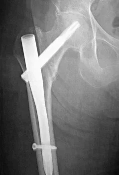
Intramedullary nail
Fixed angle plate
surgeon preference
associated femoral neck fracture
narrow medullary canal
pre-existing femoral shaft deformity

Fixed angle plate

Nail