Calcium hydroxyapatite crystals are deposited near the rotator cuff insertion,with the possibility of tendon degeneration
associated with subacromial impingement
typically affects patients aged 30 to 60
more common in women
Presents as a subacromial impingement, sometimes capsulitis
Can be associatied with endocrine disorders
diabetes
hypothyroidism
Classification
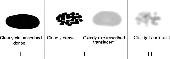
Gartner and Heyer Classification
Mole et al. Classification
A calcification dense homogenous with clear contours
B calcification dense split/separated with clear contours
C calcification non-homogenous serrated contours
D calcification dystrophic calcification of the insertion in continuity with the tuberosity
Imaging
Xray AP ( 3 rotations) + L
Internal rotation view shows infraspinatus and teres minor calcification
external rotation view shows subscapularis calcification
Attention: calcifications may be present, at the same time in differents tendons (not the same stage)
Scan : not useful
MRI : to assess rotator cuff tear
Ultrasound: to assess the limit of the calcification : deposits are hyperechoic. May also be helpful for injection and decompression
Calcification of the Biceps, supraspinatous and subscsapularis
Treatment
NSAIDs, Steroid injection, physical therapy : first line of treatment
extracorporeal shock-wave therapy : especially in the formative and resting phases
ultrasound-guided needle lavage vs. needle barbotage : especially in the resorptive stage
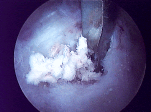
surgical decompression of calcium deposit : for refractary cases. risk of capsulitis


